Key to the successful outcome of a dental implant is the surgical procedure in which it is placed. Since the dental implant itself is merely the foundation on which a dentist will construct a crown or an attachment point for a denture; the depth, angulation, and three dimensional positioning of the implant are vital when striving to achieve the same mechanical properties nature imparts to natural teeth.
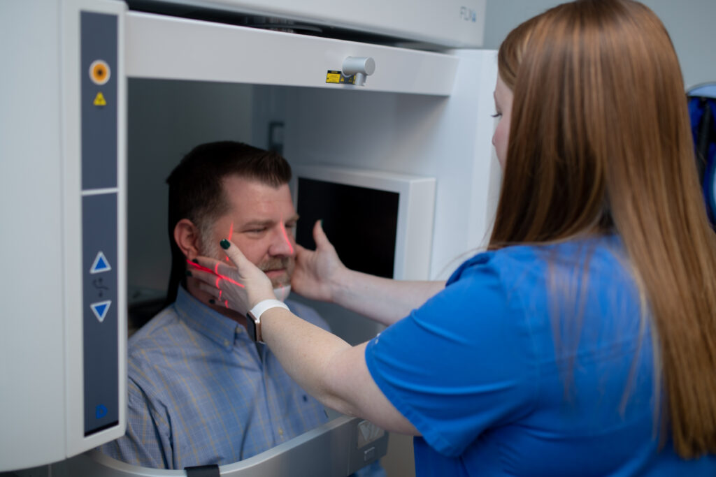
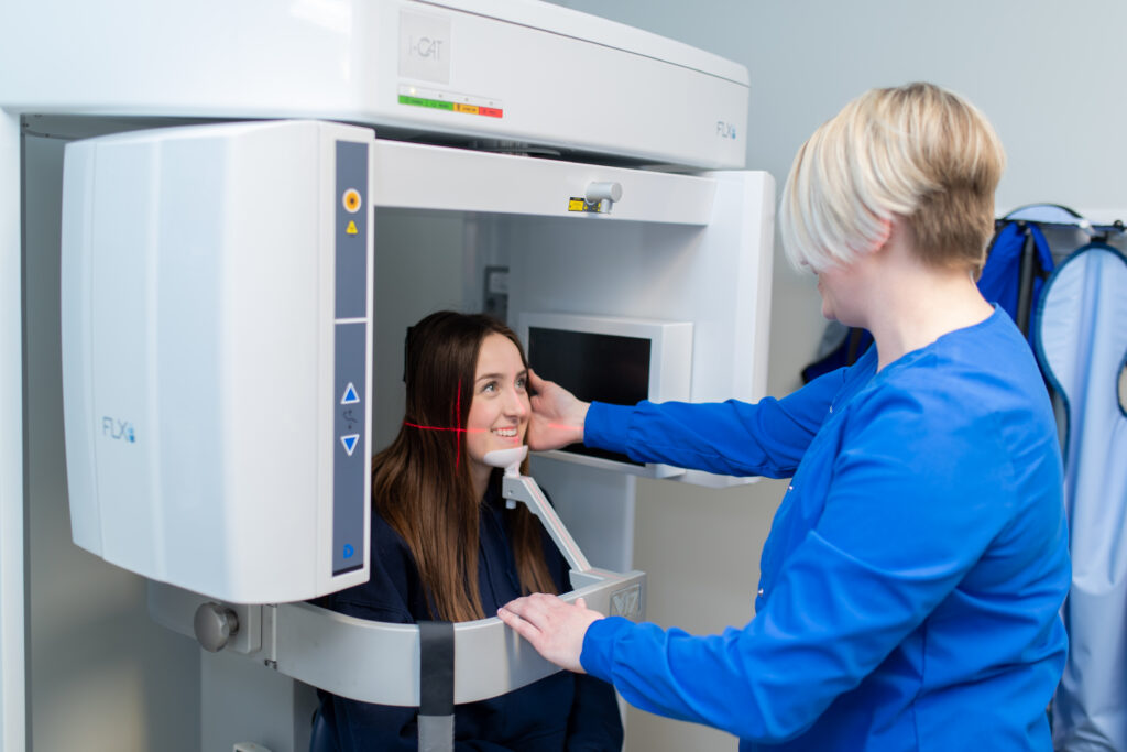
The use of three dimensional imaging represents the latest technological quantum leap in dental implant surgery. CT scans have long represented the gold standard when it comes to imaging of bone. With the advent of Cone Beam CT technology, CT scans can now be obtained comfortably and affordably in our office with much lower radiation exposure to the patient. Cone Beam CT scans can produce higher resolution images while exposing the patient to 1/10 the amount of radiation as a traditional hospital CT scanner. In addition, the CBCT machines are quieter and more comfortable, with images being acquired while the patient is seated upright rather than lying on a hard flat table.
Using specialized software, our surgeons – Dr. Busch, Dr. Otte, and Dr. Schroeder – will precisely plan the implant placement procedure on the 3D image of the patient’s facial skeleton. The 3D rendering of the patient’s jaw can be manipulated in a 360 degree environment on a computer offering a perspective that simply cannot be obtained clinically. Within the software, our surgeons will select the appropriate size, length, and manufacturer of dental implant needed and place that implant within the digital rendering of the patient’s jaw. Our surgeons can adjust the depth, angulation, and 360 degree positioning of the implant until the position is precisely where it should be to accomplish the function of the missing tooth it is replacing. In addition, the patient’s dentist, working in concert with our surgeons, can now preview the proposed positioning and even place a digital mock-up of the dental crown on the digital implant to ensure that the surgical positioning will yield an acceptable restorative result.
Once the digital planning process is complete, the data is uploaded to a specialized dental laboratory who fabricates a guide that our surgeons will use during the clinical procedure. This surgical guide allows our surgeons to duplicate the result previously planned within the computer software. Due to the precision of this process, the surgery is much less traumatic, takes less time, and has a much lower rate of complication.
At Associated Oral & Maxillofacial Surgeons, we know how to help you. Our board-certified surgeons and expert staff can provide the care you need to relieve the discomfort you feel.
"My experience at AOS was very positive. From the moment I was greeted at the door by reception to the helping caring staff, nurses, doctors and the understanding business office I felt like and individual not just a body."
"Extremely professional and great to work with. Top notch care and skilled folks from the business counter to the chair. Would highly recommend."